Pathology Poster – Lyme Disease (Lyme Borreliosis)


Lyme disease or Lyme borreliosis, a tick-borne disease, is caused by Borrelia burgdorferi.

It comprises a wide spectrum of symptoms affecting skin, musculoskeletal system, heart, eye, central and peripheral nervous system.

Visit – Pathology of Lyme Disease (Lyme Borreliosis)


lyme


October2017

Visit : Pathology Magazine – October 2017 Issue

Pathology Poster – Chikungunya


Chikungunya virus is a togavirus belonging to the genus alphavirus, indigenous to tropical Africa and Asia, where it is transmitted to humans by the bite of infected mosquitoes, usually of the genus Aedes.

Visit my website – Pathology of Chikungunya Virus Infection


chikun


October2017

Visit:  Pathology Magazine – October 2017 Issue

Pathology Poster – Onchocerciasis (River Blindness)


The filarial nematode that causes onchocerciasis is known  as Onchocerca volvulus.

Geographic distribution:  The disease is seen in  west and central Africa and in circumscribed areas of Mexico, Guatemala, Venezuela and Columbia and in parts of Yemen.

Visit :  Pathology of Onchocerciasis (River Blindness)


riverblindness


October2017

Visit – Pathology Magazine – October 2017 Issue

Coccidioidomycosis [Pathology Infographic]

Coccidioidomycosis was first described in 1892 by Alejandro Posadas (a medical student from Argentina), when he evaluated Domingo Ezcurra, a soldier who presented with a verrucous papule on the right cheek. A diagnosis of mycosis fungoides with psorospermia was considered. After having observed spherical organisms with a double-refractile outer wall, that appeared similar to the protozoan Coccidia, Posadas and his mentor, Robert Wernicke, believed that these organisms had a parasitic origin. Ezcurra died in 1898 despite many topical treatments. His head is now conserved in a museum in Buenos Aires.

Ref:Garcia SCG, Alanis JCS, Flores MG, Gonzalez SEG, Cabrera LV, Candiani JO. Coccidioidomycosis and the skin: a comprehensive review. Anais Brasileiros de Dermatologia. 2015;90(5):610-619. doi:10.1590/abd1806-4841.20153805.

Visit: Pathology of Coccidioidomycosis


coccido


Educational Gif – Pathology of Respiratory Epithelial Adenomatoid Hamartoma


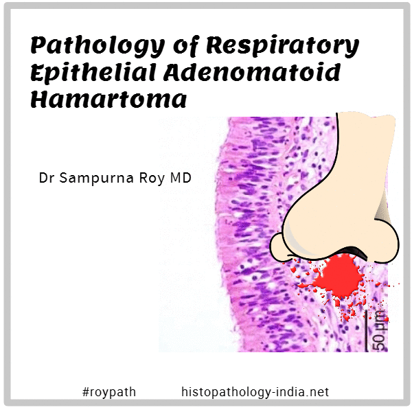
REAH is an expansile mass that causes upper respiratory symptoms and discomfort mainly in adults .

Symptoms at presentation vary and are similar to those that accompany chronic sinusitis, eg, nasal congestion, nasal obstruction, headaches, facial pain, epistaxis, and hyposmia.

Endoscopy does not reveal any distinguishing features to suggest a diagnosis of REAH, and neither CT nor magnetic resonance imaging produces a specific signal intensity that can help the clinician distinguish REAH from other sinus lesions.

Distinctive histologic features of REAH include a glandular component that originates in the overlying surface respiratory epithelium and polypoid growths that represent a proliferation of respiratory epithelial adenomatoid tissue.

It is important to recognize this lesion, however, because it can be confused histopathologically with other disease processes that require a significantly different treatment approach.

Included in the microscopic differential diagnosis of REAH are inflammatory polyps , inverted Schneiderian papillomas , and well-differentiated adenocarcinoma .

Pathologists must be aware of this entity to avoid overdiagnosis and overly aggressive surgical procedures.

GIFMaker.org_AQrOzJ
September2017