8 medical stories that touched my heart – A Pathologist

groupfriend1

Do doctors make New Year’s resolution?   We have already taken an oath to serve the patients and our community to the best of our ability.  I will continue to be a lifelong learner, apart from that I have not made any new resolution for 2016. In this post I want to share 8  medical stories that touched my heart.

doctor

  • Dr A  was a compassionate paediatrician who lost her husband and her only child in a plane crash. She was devastated and had a mental breakdown. Soon she picked herself up and vowed to turn her life around after the tragedy. Now she is a renowned paediatrician and has dedicated her life to treating children. Whenever she feels sad, she looks at dozens of little sick children depending on her. Her profession healed her own pain.

doc1

  • Dr B was a famous, charming physician and a skiing champion. He was engaged to get married to a gorgeous woman who was impressed by his outstanding skiing skills. He had a car accident and his legs were amputated. He was able to walk only with his prosthetic legs but needed a cane to support him . He didn’t want to be a burden to anyone. He left his city and handed over his established medical practice to a friend. He started working in a remote hospital in a small town. He saved lives of many poor patients. His fiancée traced him to the little town and confessed that she loved him, not his skiing skills. They got married and lived happily ever after in the little town. Sometimes they look at the skiing photographs and laugh together.

doc66

  • Dr C was a dedicated surgeon who was often on call in the night to perform emergency operations. She was successful in every operation at a young age. Her lifelong mentor and teacher instructed her to operate on a very complicated case. It was a lost case, but she made a desperate attempt to save the patient. Her mentor wanted to teach her an important lesson. He explained to her that however hard a doctor can try, some patients cannot be saved. The man she loved had terminal cancer and her husband committed suicide because he did not trust her. Once again her chosen profession saved her from mental breakdown. Her mentor trained her to be an expert surgeon. She got a national award for lifelong dedication to her work. She dedicated the rest of her life to her profession.

doc2

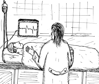
  • Dr D was a renowned gynaecologist and delivered hundreds of healthy babies. He was particularly well-known for competently handling complicated gynaecological cases. His wife was pregnant. He was doing the follow up. There were signs of complications and he was concerned. He was attending a complicating, emergency case. His wife suddenly started having severe premature labour pain. Her husband could not be reached in the operation theatre.  By the time he successfully saved the life of a mother and child, his own wife and child had died.

girlreading

  • Dr E was a bubbly, cheerful haematologist. She often felt tired at work, sometimes had mild fever and headache. Doctors are bad patients. She ignored the ominous symptoms.  Later she casually examined her blood sample and was shocked at the findings of her own blood smear. She had acute lymphocytic leukemia. It is also known as acute lymphoblastic leukaemia, which commonly occurs in children. She was particularly surprised because it is very rare in adults. Later bone marrow biopsy confirmed the diagnosis. There was complete remission within 5 weeks of starting treatment. Now she is writing a book and spending more time with her family.

doc3

  • Dr F was a pathologist who was rushing to his lab and his car collided with another car. Both his arms were bruised and blood was oozing from multiple ulcerated areas. He did not take sick leave. After preliminary treatment he went back to his lab and reported all the pending urgent cases. Other doctors were on vacation.
  • doc2  doc1
  • Dr G was a honest and sacrificing doctor. He was an orphan and was brought up by a wealthy family who treated him like their own son Dr H.  A time comes when Dr G is in a situation where he had to give up his love and profession for the family who gave him so much in life.
    Dr G and Dr H loved each other like real brothers.
    Dr H married the girl Dr G secretly loved. Dr H started medical practice and charged exorbitant amount of money from patients, especially to do illegal abortions. Dr G tried to stop him but Dr H did not listen to him. One day a patient died during illegal abortion and Dr H  was arrested. Dr G tells the police that he was the real culprit and changed all the hospital records to prove his point. In jail Dr G requested Dr H and his beloved wife to use medical profession to serve people, not to earn money. They kept the promise and  started working in a small primary health centre in a remote village. Dr G was released from jail after 9 years. His foster brother Dr H and his wife were waiting for him, with their little child.

doc5

  • Dr I  was a honest and committed oncologist. He met a patient suffering from malignant tumour of the bowel. Doctor was charmed by the charismatic patient at the first meeting. The patient knew about the seriousness of the condition. He wanted to live life to the fullest before the inevitable occurred. He touched the lives of everyone he met. He brought happiness, laughter and joy in the personal life of the serious doctor. Finally he dies laughing. The doctor broke down and cried for the patient who became his dearest friend. Dr I wrote a book from his own diary about his amazing friend and made him immortal.

“In the end, it’s not the years in your life that count. It’s the life in your years. Abraham Lincoln

Happy 2016 to all my readers.


Raining down appearance of Pigmented Spindle Cell Nevus (Reed Nevus)

Pigmented Spindle Cell Nevus (Reed Nevus) is a distinct pigmented tumour of the skin.  Clinically, this lesion presents as symmetrical, sharply circumscribed, darkly pigmented nodule or papule.  It is usually noted on the extremities and on the back. This lesion should not be mistaken for a melanoma.

Reed


Do you know why Rudolph the Reindeer has a red nose? Ask your Pathologist!

Rudolph the rednosed reindeer had a very shiny nose. And if you ever saw it, you would even say it glows.”

reindeer

Everyone has seen pictures of reindeer, for they are the animals that are said to draw the sledge of Father Christmas. Reindeers are protected from the cold by a thick skin and two coats of hair, a long coarse outer one and a fine, woolly inner one. The colour of the outer coat changes from dark brown in summer to a lighter brown in winter.

Reindeer in Norwegian arctic region has a distinct pink coloration at tip of nose.

Can Ince et al studied the functional morphology of the nasal microcirculation in humans in comparison with reindeers. (Why Rudolph’s nose is red: observational study –BMJ 2012;345:e8311).

reindeer2

The nasal microcirculation has important roles such as heating, filtering, and humidifying inhaled air, controlling inflammation, transporting fluid for mucous formation, and delivering oxygen to the nasal parenchymal cells.

The pathophysiology of many nasal conditions, such as congestion and epistaxis, is based on the regulatory mechanisms of the microcirculation.

Nasal mucosa has an important part in the uptake of drugs and responses to allergens.

The introduction of hand-held intravital video microscopes has helped in direct visualisation of the nasal microcirculation in humans.

These imaging instruments have had a special impact on intensive care medicine as they have shown the nasal microcirculation to be the most sensitive haemodynamic indicator of outcome and response to treatment. These instruments have also identified the microcirculation as a key factor in a wide range of other diseases, including diagnostic support and treatment responses in oncology.

reindeer1

Using the technique of hand-held vital video microscopy the authors characterised the microvasculature of the human nose and applied the same technique to reindeer for comparative purposes.

Intravital video microscope showed  complex architecture of the nasal microcirculation, including the kinetics of flowing red blood cells, and provided new insights into the adaptive behaviour of vascular structures under varying clinical conditions.

An interesting finding was the presence of gland-like structures in the nasal mucosa. The  explanation for the function of these circular vascular structures is mucous secretion. These structures are scattered throughout the nose and maintain an optimal nasal climate during humid weather and extremes of temperature as well as being responsible for fluid transport and acting as a barrier.

reindeer4

The nasal microcirculation in humans consists of hairpin-like vessels, microcirculatory networks, and crypt-like structures surrounded by capillaries.

“The microcirculation of the nasal mucosa in reindeer is richly vascularised and 25% denser than that in humans. These factors explain why the nose of Rudolph, the lead flying reindeer employed by Santa Claus to pull his sleigh, is red and well adapted to carrying out his duties in extreme temperatures. ”

reindeer3


Why Rudolph’s nose is red: observational study –BMJ 2012;345:e8311
 clipart courtesy: www.clipartlord.com ;  cartoon: www.toondoo.com


Standing under the Mistletoe? It is an anticancer agent


What does Christmas remind you of ? Mistletoe, holly, Christmas tree, Christmas pudding, and so many wonderful things. When the heart is happy, Christmas can never be dull. It’s not just about feasting and fun. It’s also a time for happy reunions with family and friends. 

holly     holly

Mistletoe is known to bring happiness, safety, and good fortune as long as it does not touch the ground. This is probably why it is hung up in homes in Christmas time and is supposed to bring luck to those who kiss under it.

Extracts and preparations from the parasitic plant mistletoe (Viscum album ) have been used in the treatment of cancer for decades.

First recorded use in oncology was by the Dutch physician Ita Wegman who used a mistletoe extraction for the treatment of a breast cancer patient following a recommendation by Rudolf Steiner in 1917.

mistletoe

Extracts from the plant are used in adjuvant cancer therapy mainly as injections.

The most important active agents are lectins, which have cytotoxic and immunostimulating effects.

Mistletoe extracts have low toxicity. No fatal side effects have been reported.

Breast cancer is among the most frequent types of cancer in women worldwide. Current conventional treatment options are accompanied by side effects. Mistletoe is amongst the important herbal medicines traditionally used as complementary remedies.

The benefit of mistletoe in laryngeal cancer treatment requires further investigation, and might be considered in selected patients, as an adjunct or when other conventional therapies have failed.

mistletoe1

Mistletoes of the Loranthaceae and Viscaceae are hemiparasitic plants and their preparations in the form of injectable extracts, infusions, tinctures, fluid extracts or tea bags are widely used in various cultures in almost every continent to treat or manage various health problems including hypertension, diabetes mellitus, inflammatory conditions, irregular menstruations, menopause, epilepsy, arthritis, cancer, etc.

In Germany mistletoe extract is one of the most commonly used complementary therapeutic strategies in the treatment of urological tumours.

Clinical effects of mistletoe products include improvement of quality of life, reduction of side effects from chemotherapy and radiation, and possibly increased survival.

In central Europe, white-berried mistletoe (Viscum album) preparations not only are among the most common types of treatments used in integrative medicine but also have been among of the most commonly prescribed cancer treatments in Germany per se in 2010.

By 2017, mistletoe preparations will have been used in the treatment of cancer patients for 100 years.

holly    holly


Further reading

  • [Mistletoe in the treatment of cancer].
  • Preclinical and clinical effects of mistletoe against breast cancer
  • [Mistletoe (Viscum album) preparations: an optional drug for cancer patients?].
  • Mistletoe: from basic research to clinical outcomes in cancer and other indications
  • clipart courtesy : clipartlord.com

Think before you drink – Beware of Holiday Heart Syndrome [Pathology Infographic]


Everyone was in a festive spirit. He was a healthy 50 year old man  dancing,  drinking and enjoying with his friends. One glass of wine was not enough. He consumed many glasses of alcohol  in a short span of time. Suddenly he started sweating, complained of cardiac pain, severe palpitations and respiratory distress. Finally he lost  consciousness and collapsed on the floor.

What happened to him ? 

HHS

Locally Aggressive Adnexal Tumour – Microcystic Adnexal Carcinoma [Pathology Infographic]

A slow growing locally aggressive adnexal tumour  presented as a plaque on the cheek of an elderly male.

 Just on Hematoxylin and eosin stain search for ductal structures, perineural invasion and deep invasion into the subcutaneous fat.

Immunohistochemistry will finally establish the diagnosis. Pathology of Microcystic Adnexal Carcinoma


microadntum


Leonardo Da Vinci -When Art meets Medicine


portrait
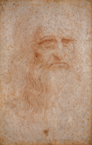
“Science comes by observation, not by authority” Leonardo Da Vinci

Presumed Self-portrait

Leonardo Da Vinci – 1452- 1519

shoulder

Leonardo Da Vinci : Superficial anatomy of the shoulder and neck (recto) –  1510  image courtesy: wikimedia-commons
Image Courtesy – wikimedia commons

Leonardo Da Vinci,  son of Caterina, a peasant girl and Ser Piero da Vinci, a Florentine notary, is one of the most amazing men who have ever lived. He is considered to be the greatest geniuses of the Italian renaissance. Not only was he a great painter, whose pictures the “Mona Lisa” and “The Last Supper” are among the most famous in the world, but also a sculptor, an architect, a poet and a composer music.

He was a genius of another kind too, for he was a pioneer in science, he studied mathematics and engineering and his ideas and the discoveries he made in work were hundreds of years in advance of his time.

His studies of human anatomy was based on meticulous dissection and showed great knowledge which was far superior to his contemporaries in medical profession.

According to Dr Kenneth D Keele’s strong words -” thus in anatomy as in so many aspects of life’s work, Leonardo was a man who awoke too early in the dawn of the Scientific Renaissance whilst others still slept”

He is an inspiration to many doctors and several interesting medical articles have been written analysing his exquisite  illustrations of human anatomy.

To his remarkable knowledge was added his skill unique skill in recording his findings in paper. These are not only important scientific diagrams but also superb works of art.

The studies of the skeleton and muscles completed between 1508 and 1510 are among his most impressive and beautiful drawings. These beautiful illustrations emphasize the fact that a good diagram will tell more than a thousand words.

The Spine (1508 -10) shows the beautiful natural curve of the vertebral  column, with the vertebrae of each part correctly numbered for the first time.

His drawing “Superficial muscles and veins of  arms shows also the superficial veins of the chest and abdomen.

embryo

Leonardo Da Vinci : Studies of Embryos by Leonardo da Vinci (Pen over red chalk 1510-1512) Image courtesy : wikimedia-commons

Leonardo Da Vinci (1452–1519) and reproductive anatomy: Arch Dis Child Fetal Neonatal Ed 1997;77: Peter M Dunn – 
He was the first to observe and comment on the fetal membranes—the chorion,  amnion, and allantois.  His drawing of a fetus in the womb is altogether remarkable and was not bettered for more than two centuries. Of that drawing, he wrote:

“In the case of this child the heart does not beat and it does not breathe because it lies continually in water. And if it were to breathe it would be drowned, and breathing is not necessary to it because it receives life and is nourished from the life and food of the mother.

The navel is the gate from which our body is formed by means of the umbilical vein… This umbilical vein is the origin of all the veins of the creature that is produced in the matrix, and it does not take its origin in any vein of the pregnant woman, because each of these veins is entirely separated and divided from the veins of the pregnant woman, and the veins and arteries are found together in pairs; and it is extremely rare for one to be found without the other being in company with it, and the artery is almost always found above the vein because the blood of the artery is the passage for the vital spirit, and the blood of the veins is that which nourishes the creature…. (the unborn child) is nourished from the same cause as the other members of the mother, and its vital powers are derived from the air which is the common living principle of the human race and other living things.”

leonardo3

Leonardo da Vinci: The Proportions of the Human Figure (Vitruvian Man) – (1490; Pen, ink and watercolour over metalpoint)Image courtesy: wikimedia-commons


leonardo4

Leonardo Da Vinci : Anatomy of the Neck – 1515 Image Courtesy – wikimedia-commons

In the course of dissection of the skull and brain Leonardo made many discoveries like maxillary and frontal sinuses (1489).

He was interested in ophthalmology and was probably first to draw the optic chiasma and to demonstrate the path of vision from the back of the eyeball to the base of the brain as in the “Optic Chiasma and cranial nerves”  (1506 -1508).

Leonardo Da Vinci described eye as the “window of the soul.”

leonardo5

Leonardo Da Vinci : Studies of human skull ( Pen, ink and black chalk on paper – 1489) Image Courtesy – wikimedia-commons 

leonardo6

In the 1490s, Leonardo Da Vinci began to investigate the heart and its work. This sketch was part of his study of the human anatomy and the circulation system. Image Courtesy – wikimedia-commons

leonardo7

Leonardo Da Vinci : Study on the proportions of head and eyes – pen and ink – second half of 15th century. Image Courtesy – wikimedia-commons

leonardo8

Leonardo da Vinci the anatomist” from McMurrich. Image Courtesy – wikimedia-commons

leonardo10

The Principle Organs and Vascular and Urino-Genital Systems of a Woman  (c. 1507) is a drawing by Leonardo da Vinci. Image Courtesy – wikimedia commons

leonardo11

View of a Skull (c. 1489) is a drawing by Leonardo da Vinci   Image Courtesy – wikimedia commons

leonardo12

Leonardo da Vinci, Drawing of the comparative anatomy of the legs of a man and a dog. Image Courtesy – wikimedia commons

Leonardo Da Vinci (1452–1519) and reproductive anatomy: Arch Dis Child Fetal Neonatal Ed 1997;77: Peter M Dunn 
He believed strongly in health care. His instructions have a remarkably modern ring:

If you would keep healthy, follow this regimen: do not eat unless you feel inclined, and sup lightly; chew well, and let what you take be well cooked and simple. He who takes medicine does himself harm; do not give way to anger and avoid close air; hold yourself upright when you rise from table and do not let yourself sleep at midday. Be temperate with wine, take a little frequently, but not at other than the proper meal-times, not on an empty stomach; neither protract nor delay the (visit to) the privy. When you take exercise let it be moderate. Do not remain with the belly recumbent and the head lowered, and see that you are well covered at night. Rest your head and keep your mind cheerful; shun wantonness, and pay attention to diet.

“As a well spent day brings happy sleep, so life well used brings happy death.” Leonardo da Vinci


[Pathology Infographic]- A Slow growing malignant tumour- Aggressive Digital Papillary Adenocarcinoma


Aggressive digital papillary adenocarcinoma is a slow growing, painless malignant sweat gland tumour. It is a rare life-threatening malignant tumour  and the diagnosis is easily missed or delayed. Both clinician and pathologist should keep this rare condition in mind when dealing with  tumours of the digits.

It is usually located on the fingers.  The tumour enlarges over several years.  Patients are usually elderly male (50 -70 year old). The clinical presentation is often confusing and can be easily misdiagnosed  for a benign lesion  such as a ganglion cyst, pyogenic granuloma, or soft-tissue infection.

Microscopic diagnosis is also challenging and it can be wrongly diagnosed as a metastatic papillary adenocarcinoma originating in the colon, thyroid, or breast. Clinicopathological correlation is  essential to rule out a possible risk of metastatic carcinoma of the skin.

These are aggressive lesions which can often recur, metastasize to distant sites, and invade local tissues including bone.

Aggressive treatment like amputation of the finger is recommended for this aggressive tumour.


dpainfo