
Alveolar soft part sarcoma is a slow growing but, highly malignant tumour.
This rare, distinctive sarcoma, typically occurring in young patients was first described by Christopherson et al in 1952.


Do doctors make New Year’s resolution? We have already taken an oath to serve the patients and our community to the best of our ability. I will continue to be a lifelong learner, apart from that I have not made any new resolution for 2016. In this post I want to share 8 medical stories that touched my heart.


“In the end, it’s not the years in your life that count. It’s the life in your years. Abraham Lincoln
Happy 2016 to all my readers.
“Rudolph the red–nosed reindeer had a very shiny nose. And if you ever saw it, you would even say it glows.”

Everyone has seen pictures of reindeer, for they are the animals that are said to draw the sledge of Father Christmas. Reindeers are protected from the cold by a thick skin and two coats of hair, a long coarse outer one and a fine, woolly inner one. The colour of the outer coat changes from dark brown in summer to a lighter brown in winter.
Reindeer in Norwegian arctic region has a distinct pink coloration at tip of nose.
Can Ince et al studied the functional morphology of the nasal microcirculation in humans in comparison with reindeers. (Why Rudolph’s nose is red: observational study –BMJ 2012;345:e8311).

The nasal microcirculation has important roles such as heating, filtering, and humidifying inhaled air, controlling inflammation, transporting fluid for mucous formation, and delivering oxygen to the nasal parenchymal cells.
The pathophysiology of many nasal conditions, such as congestion and epistaxis, is based on the regulatory mechanisms of the microcirculation.
Nasal mucosa has an important part in the uptake of drugs and responses to allergens.
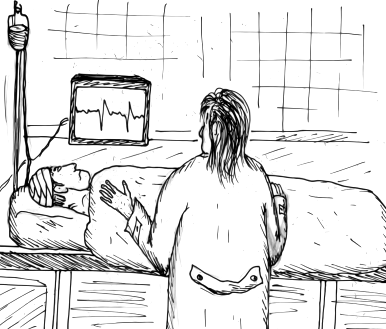
The introduction of hand-held intravital video microscopes has helped in direct visualisation of the nasal microcirculation in humans.
These imaging instruments have had a special impact on intensive care medicine as they have shown the nasal microcirculation to be the most sensitive haemodynamic indicator of outcome and response to treatment. These instruments have also identified the microcirculation as a key factor in a wide range of other diseases, including diagnostic support and treatment responses in oncology.

Using the technique of hand-held vital video microscopy the authors characterised the microvasculature of the human nose and applied the same technique to reindeer for comparative purposes.
Intravital video microscope showed complex architecture of the nasal microcirculation, including the kinetics of flowing red blood cells, and provided new insights into the adaptive behaviour of vascular structures under varying clinical conditions.
An interesting finding was the presence of gland-like structures in the nasal mucosa. The explanation for the function of these circular vascular structures is mucous secretion. These structures are scattered throughout the nose and maintain an optimal nasal climate during humid weather and extremes of temperature as well as being responsible for fluid transport and acting as a barrier.

The nasal microcirculation in humans consists of hairpin-like vessels, microcirculatory networks, and crypt-like structures surrounded by capillaries.

What does Christmas remind you of ? Mistletoe, holly, Christmas tree, Christmas pudding, and so many wonderful things. When the heart is happy, Christmas can never be dull. It’s not just about feasting and fun. It’s also a time for happy reunions with family and friends.

Mistletoe is known to bring happiness, safety, and good fortune as long as it does not touch the ground. This is probably why it is hung up in homes in Christmas time and is supposed to bring luck to those who kiss under it.
Extracts and preparations from the parasitic plant mistletoe (Viscum album ) have been used in the treatment of cancer for decades.
First recorded use in oncology was by the Dutch physician Ita Wegman who used a mistletoe extraction for the treatment of a breast cancer patient following a recommendation by Rudolf Steiner in 1917.

Extracts from the plant are used in adjuvant cancer therapy mainly as injections.
The most important active agents are lectins, which have cytotoxic and immunostimulating effects.
Mistletoe extracts have low toxicity. No fatal side effects have been reported.
Breast cancer is among the most frequent types of cancer in women worldwide. Current conventional treatment options are accompanied by side effects. Mistletoe is amongst the important herbal medicines traditionally used as complementary remedies.
The benefit of mistletoe in laryngeal cancer treatment requires further investigation, and might be considered in selected patients, as an adjunct or when other conventional therapies have failed.

Mistletoes of the Loranthaceae and Viscaceae are hemiparasitic plants and their preparations in the form of injectable extracts, infusions, tinctures, fluid extracts or tea bags are widely used in various cultures in almost every continent to treat or manage various health problems including hypertension, diabetes mellitus, inflammatory conditions, irregular menstruations, menopause, epilepsy, arthritis, cancer, etc.
In Germany mistletoe extract is one of the most commonly used complementary therapeutic strategies in the treatment of urological tumours.
Clinical effects of mistletoe products include improvement of quality of life, reduction of side effects from chemotherapy and radiation, and possibly increased survival.
In central Europe, white-berried mistletoe (Viscum album) preparations not only are among the most common types of treatments used in integrative medicine but also have been among of the most commonly prescribed cancer treatments in Germany per se in 2010.
By 2017, mistletoe preparations will have been used in the treatment of cancer patients for 100 years.
Further reading

It is absurd to divide people into good and bad. People are either charming or tedious. ~Oscar Wilde
Charisma is not so much getting people to like you as getting people to like themselves when you’re around. ~Robert Brault


Tail of the Pancreas is narrow and usually lies in contact with the inferior part of the gastric surface of the spleen. It is present within the two layers of the splenorenal ligament together with the splenic vessels, to which it is closely related.

Epididymis consist of a tortuous canal which forms the first part of the efferent route from the testis. It consists of central region, or body, a superior enlarged head, and an inferior pointed end, the tail. The lateral surfaces of the head and tail of epididymis are covered by the tunica vaginalis and hence free.

The Superolateral part of Breast is prolonged upwards and laterally towards the axilla, forming the axillary tail, which pass through the deep fascia to lie in close relationship to the pectoral group of axillary lymph nodes.

The cartilage of auricle of the external ear consists of a single piece of elastic fibrocartilage. This cartilage is absent from the lobule and between tragus and beginning of helix. Anteriorly, where the helix bends upwards, there is a small cartilaginous projection – spine of the helix. At its other extremity the cartilage is extended inferiorly as the tail of the helix. The tail of the helix is separated from the antihelix by the fissure antitragohelicina.

The caudate nucleus is an arcuate mass of gray matter which has a head, body and tail. The anterosuperior end is massive and termed head. At the interventricular foramen this narrows into body of the nucleus which tapers imperceptibly into the tail.

Dentate gyrus is a crenated strip of cortex. Anteriorly, the dentate gyrus is continued into the notch of the uncus, where it makes a sharp bend medially across the central part of its inferior surface. This transverse part is smooth and featureless, being termed the tail of the dentate gyrus (band of Giacomini), and it becomes indistinguishable on the medial aspect of the uncus.

Tailbone ( coccyx) is the terminal segment of the spine. It is a triangular bone that consists usually 3 to 5 rudimentary vertebrae fused together . The lower part of the filum terminale, also known as coccygeal ligament, inserts to the first segment. The coccyx is bordered anteriorly by the levator ani muscle and the sacrococcygeal ligament.

“A dog can express more with his tail in minutes than his owner can express with his tongue in hours.”—Anonymous
Let your life lightly dance on the edges of time like dew on the tip of a leaf. Rabindranath Tagore
When something wonderful happens we use phrases like “I feel like dancing” , “I could jump with joy”, and when we are angry we say “I’m hopping mad”. Do you know why we use these phrases of movement? We are responding to an instinct that is more ancient than speech or music. It is called “Dancing.”
Dancing is the oldest of the arts. Early man danced to describe what he was doing. This was even before he added simple music or songs to accompany the movements.
The urge to dance, or to express ourselves by the movement of our bodies, is so basic that it must have existed in men, even in the ancestors of men for thousands of years.
Dancing makes patterns in space and to have any real meaning requires a sense of rhythm. The body movement perform to some sort of regular beat or pulse. Rhythm is one of the most important features of dancing.
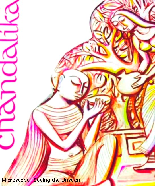
As a little child I took few simple dance lessons in the dancing school of renowned Amala Shankar. Later I learned to dance in my school Loreto House, where I studied for 12 years. Our dance teacher was famous dancer and choreographer Shanti Bose.
Along with other children I participated in the Bengali “Rabindra Nritya Natya” (Dance Drama) Chandalika. At an early age I realized that dancing was not just a matter of dance steps. Certain signs have been carefully worked out and instinctive actions play an important role.
Signs have always been used by people to communicate with each other. Example: A nod of the head means the same thing in most parts of the world. Rhythmic movement of fingers and hand gestures are used to express various emotions.
Why should children learn to dance at a young age? As a doctor now I know the reasons.
1) Dancing is an excellent form of physical exercise.
2) It is an artistic expression through the use of the body. Dancing helps in creative and artistic development of a child.
3) Dancing enhances physical function, mental health, and well-being of a toddler.
4) Dancing improves muscular endurance and strength of the legs.
5) The body becomes more flexible. There is strengthening of lower legs which improves balance and agility. Dancing increases aerobic power and makes one more energetic.
6) It psychologically prepares a child for more serious activities as it improves brain function. Learning complicated dance steps improves memory.
7) It has a positive effect on cardiovascular system and improves heart’s ability to pump blood to the lungs.
8) Dancing helps to keep the joints flexible, the muscles around the joints strong. There is greater strength of hip joint.
9) It strengthens the spinal column and the supporting muscles, ligaments, and tendons.
10) A child grows up to have a graceful bearing and gait.
11) Body of a dancer is trim and fit, with greater than average range of motion.
12) Dancing instantly elevates mood, boosts happiness, increases confidence and self-esteem and reduces stress.
As a world traveler I have seen some exceptionally beautiful dance forms like Ballet, Waltz, Tango, Belly Dancing and various types of folk dancing.
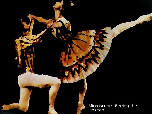
I was not born to be a dancer but I have great respect for all professional dancers. They belong to a fascinating world and will remain eternally young and healthy.

Many medical terms originate from fascinating Greek mythology. Today’s post is about a congenital malformation Cyclopia. Cyclops appear for the first time in literature in Homer’s Odyssey (8th-7th century BC).
Greek Mythology: Cyclops
In the beautiful poem called the Odyssey the Greek poet Homer describes how the hero Odysseus while returning home from Troy to his own kingdom of Ithaca, landed on an island of the Cyclopes.
Cyclopes are giants who had one eye, placed in the centre of their foreheads. They were shepherds and looked after their sheep by day, returning at night to sleep in caves.
Odyssey and some other sailors came to one of these caves which belonged to a giant called Polyphemus. The giant captured the Greeks and started to eat them for dinner.
At last Odysseus found a long pole and while Polyphemus slept, he thrust it into the eye of the giant and blinded him. Smart Odysseus told Polyphemus that his name was “Nobody”.
Brilliant idea and something to keep in mind in the modern age.
When the giant screamed out in pain other Cyclopes came running out to help him.Polyphemus cried out that “Nobody” had hurt him. To this the Cyclopes replied that if no man had hurt him then it must be one of the gods and, telling Polyphemus to bear the pain as best as he could, they went away again.
Blind Polyphemus tried in vain to catch his prisoners by feeling the back of each sheep. Odysseus and his men escaped by clinging on to the wool underneath the sheep.
In many ancient Greek stories there is evidence that one eyed Cyclopes had built great walls. The word Cyclopean is still sometimes applied to massive stone structures.
Congenital Disease – Cyclopia
Holoprosencephaly are a group of disorders arising from failure of normal forebrain development during embryonic life. There are three forms of holoprosencephaly: alobar, semilobar, and lobar varieties.
Cyclopia (alobar holoprosencephaly) is a rare and lethal human malformation, resulting from incomplete cleavage of prosencephalon into right and left hemispheres. Approximately 1 in 100,000 births are identified as infants with cyclopia.
Cyclopia typically presents with a median single eye or a partially divided eye in a single orbit, absent nose, proboscis (nose of a mammal, usually long and flexible) like structure on forehead above the eye.
Extracranial malformations described in stillbirths with cyclopia include polydactyl, renal dysplasia, and an omphalocele.
The etiology of this rare syndrome is not clear. Possible risk factors include: maternal diabetes , infections during pregnancy (TORCHs), drugs during pregnancy (alcohol, aspirin and many other teratogenic drugs), exposure to ultraviolet light, and chromosomal anomaly like trisomy 13 and genetic cause.
No one knows whether one eyed Cyclops actually existed or not, but the congenital ophthalmic disease, Cyclopia is certainly named after these mythical characters and we will never forget these giants.
Reference
Cyclopia: a rare condition with unusual presentation – a case report.
Odysseus versus the Cyclops
Cyclopia: from Greek antiquity to medical genetics.

“Today the traveller on the Nile enters a wonderland at whose gates rise the colossal pyramids of which he has had visions perhaps from earliest childhood. James Henry Breasted”.
To know the people and a country you have to visit it. Talk to the people, look into their eyes and try to understand their local culture.

The most exciting discovery of ancient Egyptian remains was that of the tomb of Tutankhamun, the last pharaoh of the XVIIIth dynasty.
The tomb was found in 1922 by an Englishman called Howard Carter who was member of an expedition team led by Lord Carnarvon. The tomb was in the Valley of the Kings, where so many Egyptian kings were buried, but this was only one that had not been broken open or robbed of its treasures.
The mummy of Tutankhamun himself was enclosed in a decorated coffin of gold, which in its turn was inside two wooden coffin and a stone one. The face of the mummy was covered with a gold mask, decorated with precious stones and there were gold ornaments on the head.
According to Dr Saleem SN et al “Subcutaneous packing procedure was used as part of mummification of royal Ancient Egyptians dated to 18th to 20th dynasties earlier than what was believed in archaeology. The Ancient Egyptian embalmers must have been skilled in dissection and possessed surgical tools that enabled them to perform this fine procedure.”
Tutankhamun became king when he was only 12 and died unexpectedly at the age of 18 years. If his tomb had not been found with all its treasure he would have been forgotten long time ago.
A curious superstition sprang up about the tomb of Tutankhamun. It was said that the people who broke it open were committing an offence against something sacred, and that they would meet sudden death as a result.
Strange incidents happened and several people connected with discovery did die tragically, one of them being Lord Carnarvon, who died in Egypt, from poisonous mosqito bite and pneumonia, five months after the finding of the tomb.
According to Dr Gandon the mysterious death of Lord Carnavon after entering the tomb of the Egyptian pharaoh Tutankhamun could be explained by an infection with a highly virulent, long living pathogenic agent. “The ‘curse of the pharaoh‘ has been used as a metaphor for the hypothesis that higher parasite propagule survival selects for higher virulence.”
The cause of King Tutankhamun’s death has been matter of speculation for several decades. Many interesting medical articles have been written on this subject.
It has been suggested that the young king was murdered by a blow to the head based on skull radiographs obtained by a team of researchers in 1968. Professor R.G. Harrison stated, “While examining X-ray pictures of Tutankhamun’s skull, I discovered a small piece of bone in the left side of the skull cavity. This could be part of the ethmoid bone, which had become dislodged from the top of the nose when an instrument was passed up the nose into the cranial cavity during the embalming process. On the other hand, the X-rays also suggest that this piece of bone is fused with the overlying skull and this could be consistent with a depressed fracture, which had healed. This could mean that Tutankhamen died from a brain hemorrhage caused by a blow to his skull from a blunt instrument.”
According to Hussein et al pathological findings included craniofacial dysmorphia, bilateral alterations of the feet, malarial disease and an acute traumatic fracture of the knee.
Hawass et al. used imaging, anatomical and DNA techniques and suggested Plasmodium falciparum malaria, concomitantly with Morbus Köhler-Freiberg, as his probable cause of death. The authors also indicated that there were no signs of gynecomastia and craniosynostoses (eg, Antley-Bixler syndrome) or Marfan syndrome. Genetic testing for STEVOR, AMA1, or MSP1 genes specific for Plasmodium falciparum revealed indications of malaria tropica in 4 mummies, including Tutankhamun‘s. These results pointed to avascular bone necrosis along with the malarial infection as the most likely cause of death of Tutankhamun.
According to Timmann et al “The age of 18–19 years attributed to Tutankhamun makes malaria as his primary cause of death rather unlikely, as a certain degree of semi-immunity may readily be assumed.” Plasmodium falciparum malaria was endemic in ancient Egypt and probably caused death. Local residents after continuous exposure to plasmodium developed immunity against both parasites and disease. Fatal courses of malaria usually affect young children (6- 9 year) and pregnant women but not adults.
According to Timmann et al bone lesions of two metatarsals of the left foot and shortening of the second left toe, indicated advanced osteonecrosis, osteomyelitis or ulcerative osteoarthritis in the article by Hawass et al. These radiographic findings are consistent with osteopathological lesions as often noted in sickle cell disease , a haematological condition described to occur in many local residents of Egypt (El-Beshlawy & Youssry 2009). The occurrence of anaemia, other pathological haematologic disorders, and of the haemoglobin mutation that causes sickle cell disease (HbS) has been demonstrated in predynastic Egyptian remains (Marin et al. 1999).
Timmann et al indicated, “in contrast to osteonecroses associated with sickle cell disease, Köhler-Freiberg disease is highly unlikely to be a co-cause of death in young adulthood.”
Avascular osteonecroses, inflamed by osteomyelitis and septicaemia due to pathogenic agents including Salmonella and Staphylococcus, are known sickle cell disease complications and may be exacerbated by malaria attacks.
Gaucher’s disease (type 1-3) should be considered which may cause painful avascular osteonecroses. Gaucher’s disease is a rare autosomal-recessive lysosomal storage disease and has been described in Egyptians.
Other features considered in the differential diagnosis of avascular osteonecroses in King Tutankhamun’s bones included other inheritable and non-hereditary disorders – trauma, sequelae after osteomyelitis, acute pancreatitis and systemic lupus erythematosus.
Brandt G. suggested the theory that Tutankhamun may have suffered from hypophosphatasia, an inherited metabolic disorder that affects especially the musculo-skeletal system in many ways. The author has compiled both medical and archaeological findings to support his theory and suggests that existing DNA samples of Tutankhamun and other members of his family should be tested for defects on the ALPL gene.
The question on Tutankhamun‘s supposed gynaecomastia cannot be answered as most of mummy’s chest wall was missing.
All the scientific findings can neither be proved or ruled out.
We do know that the most common diseases of Ancient Egypt were traumatic injuries, malaria and tuberculosis.

King Tutankhamun is one of the most famous king of Ancient Egypt. It is not surprising that extensive scientific studies have been made to find the actual cause of death. Infectious disease, metabolic disorder, neoplastic lesion, trauma (crash of his chariot causing lethal injury) and even murder have been suggested by various authors.
The cause of death of the great ruler of Egypt will continue to fascinate researchers. I wrote a similar post about Death of Alexander the great – A medical Enigma.
The difference between the two cases is that the remains of Alexander the Great was not found, hence anatomical and other scientific examinations could not be done. In case of King Tutankhamun extensive studies including virtual autopsy, imaging , anatomical and DNA analysis were performed.
Reference:
