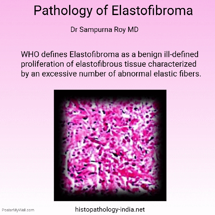
Elastofibroma is a benign slowly progressive reactive lesion involving abnormal elastogenesis.
Pathology of this soft tissue lesion is explained in an animated gif. Please visit my website for more information – Pathology of Elastofibroma.

Elastofibroma is a benign slowly progressive reactive lesion involving abnormal elastogenesis.
Pathology of this soft tissue lesion is explained in an animated gif. Please visit my website for more information – Pathology of Elastofibroma.
Eccrine spiradenomas are benign tumours , which are thought to originate from the eccrine sweat glands. They are common in young adults and are without a sex predilection. These tumours usually present as tender lesions and are typically solitary, intradermal, well-circumscribed, and firm.
Pathology of Spiradenoma is explained in the animated gif.
Please visit my website to learn more about Pathology of Spiradenoma.

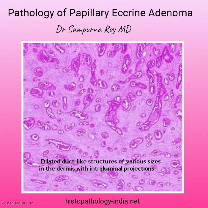
Papillary eccrine adenoma, a rare benign adnexal tumour, was first described in 1977.
Salient microscopic features are highlighted in the animated gif.
Please visit my website for more information – Pathology of Papillary Eccrine Adenoma
The rheumatoid nodule is a lesion commonly found on extra-articular areas prone to mechanical trauma.
When present with inflammatory symmetrical polyarthritis, it is pathognomonic of rheumatoid arthritis.
This educational gif briefly explains the microscopic features of a rheumatoid nodule.
Please visit my site for further information: Pathology of Rheumatoid Nodule

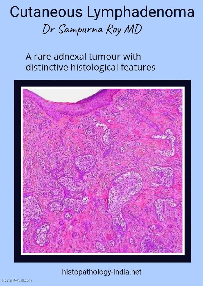
Cutaneous lymphadenoma is a rare tumour with distinctive histologic features, which was originally described as a lymphoepithelial tumour of the skin by Santa Cruz and Barr in 1987.
Pathology of cutaneous lymphadenoma is explained in one animated gif.
Please visit my website for more infomation : Pathology of Cutaneous Lymphadenoma.
Visit: Pathology Magazine – August 2017 Issue
Cylindroma is a rare skin appendage tumour that may occur either as solitary or multiple lesions.
Ancell first described multiple cylindromas of the head and abdomen in 1842.
Pathology of this appendage tumour is explained in the following educational animated gif.
For more information on cylindroma, please visit my website – Pathology of Cylindroma.

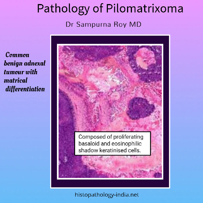
Pilomatrixoma is a rare benign skin tumor arising from multidirectional cells of hair root follicles.
It is also known as Malharbe’s calcified epithelioma.
Malharbe and Chenantais defined the lesion as calcified epithelioma in 1880 for the first time.
This animated gif explains the microscopic features of Pilomatrixoma.
For more information please visit my website: Pathology of Pilomatrixoma.
Elastosis perforans serpiginosa is a rare skin disease characterized by transepidermal elimination of abnormal elastic fibers.
Pathology of this rare skin lesion is explained in an animated gif.
Please visit my website for more information – Pathology of Elastosis Perforans Serpiginosa.

Infection with the thermally dimorphic fungus Histoplasma capsulatum can produce a wide spectrum of clinical manifestations, ranging from self-limiting respiratory complaints to progressive, life-threatening infections.
The pathology of this fungal lesion is explained in an animated gif.
For more information please visit my website: Pathology of Histoplasmosis

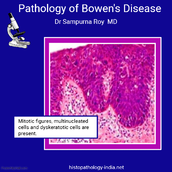
Bowen’s disease, also known as an intraepithelial squamous cell neoplasia, is a carcinoma in situ which was reported for the first time in 1912 by a dermatologist named Bowen.
Pathology of Bowen’s disease is briefly explained in this simple animated gif.
For more information please visit my website: Pathology of Bowen’s Disease